|
research
Research in Our
Laboratory
Overview of our research
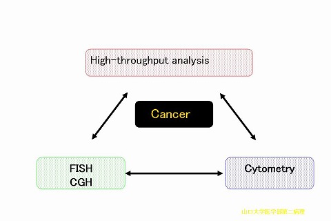
In our laboratory, we are making research with a focus on quantitave
evaluation of biological characteristics including genotype and phenotype of
human solid cancers. We employ three technologies; (1) molecular cytogenetics
such as fluorescence in situ
hybridization (FISH) and comparative genomic hybridization (CGH), (2)
high-throughput cell analysis methods including devices of cell array and
multiplex-immunostain chip, and (3) cytometry including flow cytometry and
image cytometry like laser scanning cytometry (LSC) which allows quantitative
analysis of cells. Cell array system and multiplex immunostain chips have been
originally developed in our laboratory as sophisticated cell analysis methods.
In addition, we are developing new technologies for assisting our research.
I. Cancer Molecular Cytogenetics
We are interested in genomic abnormalities of
solid tumors, which are analyzed by means of molecular cytogenetic technologies
such as fluorescence in situ
hybridization (FISH) and comparative genomic hybridization (CGH). We have
already examined more than 3,000 tumors by chromosomal CGH, and now we are
using array-based CGH as well as cromosomal CGH. We have identified genomic
alterations (copy number aberrations) linked to biological characteristics in
human solid tumors. It is indicated that CGH analysis of solid tumors provides
useful information in personalized treatment of cancer.
Topics
1. We have succeeded in developing a
gastric cancer specific mini-array allowing estimation of node metastasis,
liver metastasis, peritoneal dissemination and depth of tumor invasion. The
mini-array contains 50 BAC clones for estimating these clinicopathological
parameters. This system allows a clinicopathological state even in a single
biopsy specimen before surgical operation.

2. We have dicovered CNP (copy number
polymorphism) closely linked with cancer susceptibility (Pat. #2008-48668). The
CNP is detected in almost all patients with a specific type of cancer. CNP is
different in different type of cancer.

II. The development
of new technologies
for analysis of cellular characteristics
We have been developing new technologies with
the aim of rapid and efficient analysis of gene expression in both individual cell
and a cell population. Three representative technologies developed in this
laboratory are introduced here.
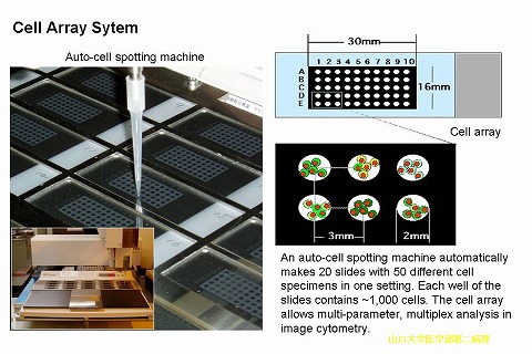
(1) Cell array
A cell array device allows analysis of cellular characteristics
such as DNA ploidy, numerical chromosomal aberrations, and antigen expression
in multiple specimens in a single experiment. Fifty (10 x 5) spots, 2 mm in
diameter, were arrayed in an area of 30 x 16 mm on a glass slide, and
approximately 1,000 cells were placed on each spot. This system is powerful in
image cytometry. (Am J Pathol 2000;157:723-8)
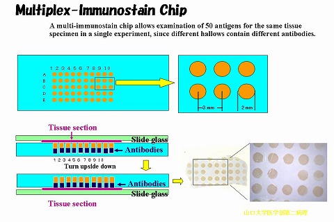
(2) Multiplex-immunostain
chip
To make immunohistochemical examination more
efficient, we have developed a novel device called a
"multiplex-immunostain chip (MI chip)." The chip is a panel of
antibodies contained in a silicon rubber plate that consists of 50 x
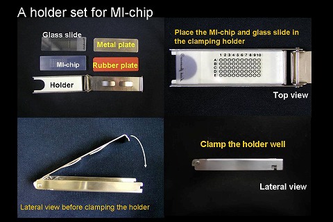
2-mm-diameter wells. A tissue section slide is placed on the plate and is
fastened tightly with a specially designed clamp. The plate with the slide is
then turned upside down, which applies the antibodies to the section. This
technology allows IHC staining of a tissue section with 50 different antibodies
in a single experiment, reducing the time, work, and expense of IHC analysis.
In addition, it enables pathologists to compare expression of multiple antigens
on a tissue section simply by changing microscopic fields on a single slide.
This device can be used in various applications in differential diagnosis of
tumors and the field of cell biology. (J Histochem Cytochem. 2004; 52: 205-10)
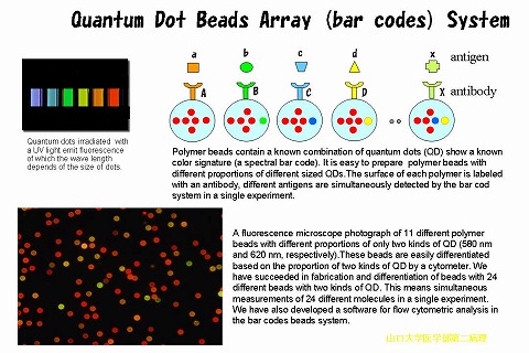
(3) Quantum dots beads array (bar codes) system
Quantum dots (QDs) show unique characteristics
as fluorescence tags. In this laboratory, we have riginally developed ‘beads
array’ consisting of beads with different proportions of two kinds of QD. We
have prepared 24 kinds of ‘bar code beads’ allowing analysis of 24 kinds of
molecules in a single experiment.
III. Cytometry
We have long experience in cytometry that is a routine technology
in biomedical research and laboratory medicine. Two types of cytometry, flow
cytometry (FCM) and Image cytometry such as laser scanning cytometry (LSC), are
routinely available in our laboratory. Both are useful for an analysis of DNA
ploidy in tumors. FCM is employed for not only cytomics but also the flow array
system, and LSC is a useful technology in the cell array system. Recently, we
have started a project concerning an application of quantum dots to cytomics,
as mentioned above.
Appendix
Virtual slide system
A virtual slide (VS) system is an interesting digital imaging tool in pathological education of medical doctors and students. The VSs that simulate real light microscopy are used in the pathological department for not only education but also telepathology. Every one can view VSs at the below web site:
http://ds.cc.yamaguchi-u.ac.jp/~2byouri/e-link.html
introduction
|