
メンバー
| チーフ | 岡村 誉之 |
|---|---|
| メンバー |
藤村 達大 宮崎 要介 末冨 建 松山 哲也 中田 祐樹 兼行 恵太 |
研究内容
当グループでは、血管内超音波法(Intravascular Ultrasound;IVUS)を用いた冠動脈内の動脈硬化病変に関する研究を長年にわたり行ってきました。これまでに、冠動脈粥腫(プラーク)の組織性状評価法の開発(Wavelet解析・Radio-frequency解析)、冠動脈イメージングの3次元化(3D-IVUS解析)、プラーク破裂機序の解明(Shear stress解析・シミュレーション解析)などを行い、その成果を国内外で発表してきました。
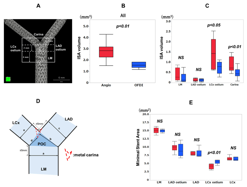
近年、IVUSよりも高解像度の光干渉断層法(Optical Coherence Tomography;OCT)が登場し、冠動脈内病変およびその治療をより詳細に評価することが可能となりました。特に、OCT画像を3次元化した3D-OCTは、病変を立体的に可視化できるだけでなく、ステントストラットの形状を正確に把握することが可能であり、冠動脈分岐部治療において有用と考えられています。当研究チームでは、冠動脈分岐部病変のステント治療における3D-OCTの有用性や、3D-OCTを用いた冠動脈分岐角度の評価、定量的冠血流予備量比(Quantitative Flow Ratio;QFR)による冠動脈狭窄に対する虚血評価、MDCTと冠動脈分岐部病変との関連性について研究を進めており、その成果を報告してきました。
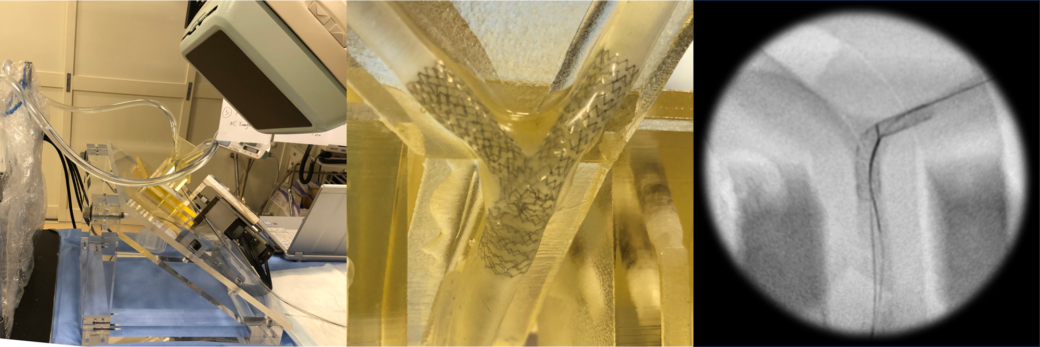
また、早稲田大学(TWINs)と共同で左冠動脈主幹部分岐部の拍動病変モデルを作成し、3D-OCT/3D-OFDIガイド下にTwo-stent留置を行い、血管内イメージングの有用性およびTwo-stent治療の特性についても研究を行っています。さらに、多施設共同研究にも積極的に参加し、調査研究および情報発信を精力的に行っています。
他施設共同研究
現在進行中
CANDO試験、3D-OCT Bifurcation Registry (冠動脈分岐部病変治療に対するOCT観察研究)、OPTIMA-AF試験、PEMA-COMA試験、YAMAMI Registry
これまで携わってきた研究
OPINION試験、ABSORB Japan、Mechanism AMI、ILUMINEN III、JAPAN-ACS、PACIFIC Registry、OPTIMUM study、3D-OCT Bifurcation Registry、Mechanism Ultimaster、ASET-JAPAN、OPTIVUS、FAVOR Ⅱ Japan
使用可能なソフトウエア
- ANSYS (数値流体解析が可能なソフトウエア)
- Medis 2D-QCA/ 3D-QCA/ Q-IVUS/ QAngio OCT/ QFR
- Strut Detector
- Realia (2D画像から3D画像作成が可能なソフトウエア)
- 3-mension (CT画像から大動脈弁および末梢血管に対し正確な情報の解析が可能なソフトウエアで主にTAVI前解析に用いられている)
- Simpleware(STL画像修正が可能なソフトウエア)
研究実績
Fujimura T, Takemitsu M, Murayama R, et al. Appropriate Selection of the Initial Diagnostic Catheter for Left Coronary Angiography Using Computed Tomography. Cureus. 2024 Dec 2;16(12): e75004.
Nakata Y, Ishiguchi H, Fujimura T, et al. Platypnea-Orthodeoxia Syndrome - Sequential Comparison of the Ascending Aorta’s Anteversion to the Right Atrium. Circ Rep. 2024 Apr 11;6(5):187-188.
Matsuyama T, Fujimura T, Okamura T, et al. Lesion of slit-like stenosis underestimated by fractional flow reserve based on computed tomography images. Int J Cardiovasc Imaging. 2024 May;40(5):1153-1155.
Akase H, Okamura T, Nagoshi R, et al. Risk Assessment of Side Branch Compromise After Coronary Bifurcation Stenting - A Substudy of the 3D-OCT Bifurcation Registry. Circ J. 2024 May 24;88(6):959-969.
Matsuyama T, Okamura T, Miyazaki Y, et al. Type 2 myocardial infarction due to cardiogenic shock in severe aortic stenosis confirmed by computed tomography and pathological analysis. Eur Heart J Case Rep. 2023 Aug 17;7(8): ytad410.
Okamura T, Iwasaki K, Lu H, et al. Importance of optimal rewiring guided by 3-dimensional optical frequency domain imaging during double-kissing culotte stenting demonstrated through a novel bench model.
Sci Rep. 2023 Aug 19;13(1):13511.
Fujimura T, Okamura T, Nagoshi R, et al. Serial changes of the side-branch ostial area after single crossover stenting with kissing-balloon inflation. Int J Cardiovasc Imaging. 2023 Aug;39(8):1593-1603.
Akase H, Okamura T, Fujimura T, et al. Mechanism of a Stuck Crown of the Orbital Atherectomy System and Successful Retrieval Procedure. JACC Cardiovasc Interv. 2022 Apr 25;15(8): e89-e90.
Nishimura T, Okamura T, Fujimura T, et al. Feasibility, reproducibility and characteristics of coronary bifurcation type assessment by three-dimensional optical coherence tomography. PLoS One. 2022 Feb 1;17(2): e0263246.
Takenaka H, Okamura T, Miyazaki Y, et al. Serial changes in the quantitative flow ratio in patients with intermediate residual stenosis after percutaneous coronary intervention. Heart Vessels. 2022 Mar;37(3):363-373.
Fujimura T, Okamura T, Furuya K, et al. Comparison of diagnostic performance in assessing the rewiring position into a jailed side branch between online 3D reconstruction systems version 1.1 and 1.2 derived from optical frequency domain imaging. CVIT 2020 Oct;35(4):336-342.
Okamura T, Nagoshi R, Fujimura T, et al. Impact of Guidewire Recrossing Point into Stent Jailed Side Branch for Optimal Kissing Balloon Dilatation – Corelab 3D Optical Coherence Tomography Analysis. EuroIntervention 2018 Feb 2;13(15): e1785-e1793.
Fujimura T, Okamura T, Tateishi H, et al. Serial changes in the side-branch ostial area after main-vessel stenting with kissing balloon inflation for coronary bifurcation lesions, assessed by 3D optical coherence tomography. EHJ Cardiovasc Imaging 2017.
Nakamura T, Okamura T, Fujimura T, et al. Serial changes in the three-dimensional aspect of the side-branch ostium jailed by a drug-eluting stent assessed by optical coherence tomography. IJC Imaging 2017.
Okamura T, Fujimura T, Yano M. Three-dimensional reconstruction of optical coherence tomography for improving bifurcation stenting. Journal of Cardiology Cases 2016;13(5):137-8.
Oda T, Okamura T, Yamada J, et al. Comparison of neointimal coverage and extra-stent lumen between sirolimus and everolimus-eluting stent using optical coherence tomography. Heart Vessels 2016;31(4):449-56.
Nakao F, Okamura T, Suetomi T, et al. Differences of side branch jailing between left main-left anterior descending artery stenting and left main-left circumflex artery stenting with Nobori biolimus-eluting stent. Heart Vessels 2016 Dec;31(12):1895-1903.
Maeda T, Okamura T, Yamada J, et al. Serial three-dimensional optical coherence tomography assessment of strut coverage and intraluminal structures after drug-eluting stent implantation. CVIT 2014;29(1):31-9.
Tateishi H, Okamura T, Yamada J, et al. Sequel of Jailed Side Branch. Circulation Journal 2014;78(3):772-4.
Okamura T, Onuma Y, Yamada J, et al. 3D optical coherence tomography: new insights into the process of optimal rewiring of side branches during bifurcational stenting. EuroIntervention 2014;10: 907-15.
Okamura T, Matsuzaki M. Sirolimus-eluting stent fracture detection by three-dimensional optical coherence tomography. CCT 2012 Mar 1;79(4):628-32.
Okamura T, Yamada J, Nao T, et al. Three-dimensional optical coherence tomography assessment of coronary wire re-crossing position during bifurcation stenting. EuroIntervention 2011 Nov;7(7):886-7.
Okamura T, Hiro T, Fujii T, Yamada J, Fukumoto Y, Hashimoto G, et al. Late giant coronary aneurysm associated with a fracture of sirolimus eluting stent: a case report. J Cardiol 2008;51(1):74-9.
Fukumoto Y, Hiro T, Fujii T, et al. Localized elevation of shear stress is related to coronary plaque rupture: a 3-dimensional intravascular ultrasound study with in-vivo color mapping of shear stress distribution. JACC 2008;51(6):645-50.
Hashimoto G, Hiro T, Matsuzaki M. [Current status and perspectives of coronary imaging for patients with diabetes mellitus]. Nihon rinsho Japanese journal of clinical medicine. 2006;64(11):2052-61.
Imoto K, Hiro T, Fujii T, Murashige A, Fukumoto Y, Hashimoto G, et al. Longitudinal structural determinants of atherosclerotic plaque vulnerability: a computational analysis of stress distribution using vessel models and three-dimensional intravascular ultrasound imaging. JACC 2005;46(8): 1507-15.
Murashige A, Hiro T, Fujii T, Imoto K, Murata T, Fukumoto Y, et al. Detection of lipid-laden atherosclerotic plaque by wavelet analysis of radiofrequency intravascular ultrasound signals: in vitro validation and preliminary in vivo application. JACC 2005;45(12): 1954-60.
Hiro T, Matsuzaki M. [Acute coronary syndrome–an overview of its pathophysiology]. Nihon rinsho Japanese journal of clinical medicine. 2003;61 Suppl 5:259-65.
Murata T, Hiro T, Fujii T, Yasumoto K, Murashige A, Kohno M, et al. Impact of the Cross-Sectional Geometry of the Post-Deployment Coronary Stent on In-Stent Neointimal Hyperplasia An Intravascular Ultrasound Study. Circulation Journal. 2002;66: 489-93.